

How DNA Evidence Is Used in Sexual Assault Cases – A Real Case Explained

DNA evidence is powerfully used to prosecute sexual assault cases in the Australian justice system. For a defendant facing DNA evidence, the process can feel daunting and highly complex.
This article explains a real case from commencement all the way up to trial, and is written by the DNA expert engaged by defence. At first glance the evidence appeared damning, with the prosecution alleging that semen matching the defendant was detected within the vagina, and that there was matching DNA. However, forensic evidence can be wrong, and DNA does not always support the alleged activities, particularly when the parties are in social contact.
Whilst court outcomes are legal, this matter and others like it, can find themselves decided on the science.
In this case, the defence team argued that the semen and DNA did not substantially support the allegations and the jury agreed in returning a not guilty verdict.
When DNA evidence becomes a problem in a caSE
DNA evidence does not automatically prove what happened.
In many sexual assault cases, the scientific results appear clear at first glance. But the real issue is not be merely whether DNA is present — it is whether the results can actually support the alleged events.
In some matters:
– DNA cannot be linked to a specific body fluid
– DNA may be consistent with social contact
– Statistics may be based on the wrong comparison
– Testing methods may not separate DNA sources properly
– Transfer and persistence may provide alternate explanations
These are not rare issues. They are the types of problems that are examined during an independent forensic review.
→ Read: When DNA statistics are wrong
→ Read: When courts refuse DNA evidence
This detailed article is ideal for a person facing sexual assault charges which rely on DNA, and for lawyers who are not closely familiar with forensic DNA expert evidence. The science is explained in terms of how DNA evidence works in criminal matters. Deeper scientific reasoning and study paper citations are provided behind the Read more scientific detail sections.
Names, times and details have been edited for privacy, whilst forensic accuracy has been maintained.
Can DNA Evidence Prove Sexual Assault?
DNA evidence does not prove sexual assault on its own. DNA may show that semen, saliva, or skin cells are present, but it cannot establish consent, timing, or how contact occurred. For this reason, DNA evidence in sexual assault cases must always be interpreted alongside the surrounding circumstances.
How a Sexual Assault Allegation Involving DNA Actually Unfolds
Many people first encounter DNA evidence through an allegation and have no prior experience with how the process works. This article explains how DNA evidence is actually interpreted in a real case, not just how it appears in a laboratory report.
It was alleged by the complainant that there was vaginal-penile intercourse and that the male also digitally penetrated her vagina with his fingers. The defendant denies the sexual activities, however, acknowledges knowing and being in social contact with the complainant.
Individuals facing allegations involving DNA evidence often assume that legal representation alone will address the scientific issues. In practice, criminal defence lawyers manage the legal process, while the interpretation of DNA evidence remains a specialised forensic task. The two disciplines operate alongside one another, but one does not replace the other.
In this matter, the social interaction between the parties was important. There was accepted, non-incriminating contact, meaning the presence of DNA alone did not automatically point to the alleged activity.
The government laboratory reported that DNA matching the defendant was detected on the complainant, along with findings interpreted as semen. However, those laboratory reports are limited in scope. They address whether biological material is present and whether DNA profiles can be obtained; they do not determine whether those results support the prosecution allegations or whether those results are equally consistent with social contact.
At an early stage, defence counsel sought an opinion from Roebuck Forensics on the weight of the scientific evidence. In his words: “is there something I’m missing in the results, or should I be advising my client to plead?”
An initial review was conducted by Ms Roebuck, which revealed significant issues and Counsel was advised. The defence formally instructed Roebuck Forensics to thoroughly examine the laboratory material in detail and determine:
- – Is the DNA result and statistic robust?
- – Is the semen result conclusive?
- – Is the DNA from the semen?
- – Do the scientific results support the prosecution allegations?
- – Do the scientific results support the defence account of social interaction?
The Role of DNA Evidence in Sexual Assault Cases
It is important to understand that the role of the government laboratory and the role of an independent forensic expert are very different.
Essentially, the crown will serve its DNA evidence, whilst not necessarily seeking to establish whether it specifically supports the allegations. What that meant in this case (and in many others) is the laboratory report set out whether DNA was detected, whose DNA it may be, and whether semen was detected – in forensic science this is known as the “source” results (addressing the “who” of DNA). The crown DNA report does not evidence consideration or opinions around “how” the DNA came to be present – this is referred to as the “activity” in forensic science.
In this matter, the central underlying question before the court, is ‘how did the DNA get there’?
As is often the case, Mr Smith and Ms Hood acknowledge prior social contact. Therefore, the mere presence of male DNA only goes so far towards addressing the central question before the court.
However, it would be dangerous to assume that reported DNA and semen is not extremely compelling to a jury. To a jury, these results can appear absolute.
This is where independent forensic review goes further. An independent expert will evaluate and explain the DNA results, and report as to what those results do and do not support. This includes examining whether mistakes have been made with the testing and whether those results appear overstated.
Further, and importantly, independent reporting will give written consideration to the relevant background information and whether the DNA results are consistent with the allegations; or consistent with alternative explanations, such as prior social contact.
Prior Social Contact Between the Parties
There is often social interaction between the parties, and the specific detail of that interaction can be highly relevant scientifically.
On the night of the alleged events, Ms Hood and Mr Smith were socialising together at a pub for a number of hours. CCTV footage captured the parties talking, drinking from beer glasses, sitting near one another, and attending the same bar counter.
On one occasion the parties returned to their seating and switched seats. There were instances of casual touching including when Mr Smith put his right arm around the waist of Ms Hood. The footage from one passageway established that the parties had each walked towards the bathrooms and returned minutes later, consistent with them each having toileted numerous times throughout the evening.
The complainant set out in her statement that she offered Mr Smith a lift home because he lived close to her. Ms Hood states that during the 15-minute drive, there was kissing and non-sexual touching. During the drive, the parties agreed to continue to Ms Hood’s home for another drink. Upon arrival, the parties sat on the living room sofa and shared some wine which was poured from a previously opened bottle into glasses. It was uncontested that further kissing, non-sexual touching and toileting occurred at the house. By Ms Hood’s account, about an hour or two after arriving, she awoke to find Mr Smith penetrating her vagina with his penis. Words were said to be exchanged, such that the complainant believed her vagina had also been penetrated digitally. The complainant was unsure if a condom was used and could not be certain that there was ejaculation.
Why Prior Social Contact Matters in DNA Evidence
We deposit our DNA onto our surroundings when we touch, sneeze, cough, sweat, bleed and even when we talk. This DNA can originate from our skin cells or from our body fluids. It can persist upon surfaces for significant periods of time, even years. This DNA can then be subsequently relocated onto other surfaces, via the process of DNA transfer. In sexual assault investigations, trace DNA may be detected even where no direct sexual contact occurred, because trace DNA can transfer through everyday interactions.
Secondary DNA transfer is a recognised phenomenon in sexual assault cases and can explain how a person’s DNA appears on an item they never directly touched. Indirect DNA transfer describes the common scenario of our DNA being upon a surface without us directly contacting that surface. For example, you could hold someone’s hand and deposit DNA onto that persons hand – then that person adjusts their clothing and deposits your DNA onto the waistband of their their underpants. The sequence can continue, when for example the same person later goes to the bathroom, and pushes their underpants downwards to urinate, they can collect your DNA again from the hips, and then deposit your DNA upon their vagina during wiping.
Indirect transfer – Secondary trnasfer – Direct transfer – DNA parking – Bi directional transfer
Read more scientific detail
DNA transfer is the reason your DNA can be found on an item you never touched.
Modern forensic science recognises that genetic material can move between people, surfaces and objects through everyday interactions, sometimes across multiple steps, before it is ever detected.
The presence of an individual’s DNA on an item is often interpreted as evidence that they must have handled it. While this was a reasonable assumption in the early years of forensic DNA testing, scientific understanding has developed substantially. Since the early 1990s, it has been known that DNA can be recovered from objects following simple handling. It is now equally well established that DNA may be present even where no direct contact occurred. This is due to the phenomenon known as DNA transfer.
DNA transfer refers to the movement of genetic material from one person to an object, or between objects, through a variety of mechanisms. These include direct transfer (through physical contact), indirect transfer (via an intermediary surface or person), and secondary, tertiary, or even quaternary transfer, where DNA is passed through multiple steps before being detected. In practical terms, this means that a person’s DNA may be detected on an item they never handled, simply because it was transferred there through everyday interactions.
Direct transfer occurs when biological material is deposited through physical contact – for example, handling an item, wearing clothing, or touching another person. This form of transfer was historically the primary assumption underlying forensic interpretation.
Indirect transfer occurs when DNA is moved via an intermediate surface or person. Secondary transfer describes DNA passing from one individual to another person or object, and then onto a final item. Tertiary and quaternary transfer extend this concept further, where DNA may pass through multiple stages before being detected. These mechanisms are not theoretical; they have been demonstrated repeatedly in controlled experimental studies and in real casework environments.
Related concepts have also emerged in the forensic literature. “DNA parking” describes the persistence of DNA on a surface after initial deposition, allowing it to be recovered later in circumstances unrelated to the original contact. “Bi-directional transfer” recognises that DNA can move in both directions when two surfaces or individuals interact – not simply from one person onto an item, but also from the item or environment back onto the person.
These processes are influenced by many variables, including the nature of the surface, the amount and type of biological material present, the duration and pressure of contact, environmental conditions, and subsequent handling. Individual differences in shedding – the extent to which a person deposits DNA – also play a role. Some individuals deposit DNA readily, while others leave little detectable material even after direct contact.
For this reason, the detection of DNA does not, in itself, establish how or when it was deposited. The presence of a DNA profile can indicate that genetic material from a person is present on an item, but it cannot determine whether that person handled the item, when any contact occurred, or whether the DNA arrived there through transfer rather than direct interaction.
In casework, this distinction is critical. DNA findings are often reported at the level of source – identifying whose DNA is present – while the forensic question before the court frequently concerns activity: what happened, and how did the DNA come to be there. The mechanisms of transfer, persistence, and background DNA mean that source attribution alone cannot resolve that question.
Scientific interpretation is therefore required to evaluate whether the observed findings are consistent with direct contact, indirect transfer, or alternative mechanisms. This involves consideration of the item involved, the circumstances alleged, the sampling strategy, the nature of the DNA profile obtained, and the broader evidential context. Without this evaluation, there is a risk that the presence of DNA will be assumed to reflect direct involvement, when the science does not support that conclusion.
The evaluation of DNA transfer has developed through extensive research over the past three decades. Work by researchers including van Oorschot, Goray, Kokshoorn, Taylor, and others has demonstrated the prevalence of indirect and secondary transfer, the variability of DNA persistence, and the complexity of interpreting trace DNA in real-world environments. These studies form the scientific foundation for modern activity-level interpretation in forensic biology.
In reviewing the evidence and forming my opinions, I rely on a substantial body of peer-reviewed literature addressing direct and indirect transfer, secondary, tertiary and quaternary transfer, persistence, shedding variability, and the interpretation of trace DNA findings in forensic casework. This research provides the framework within which DNA findings must be evaluated, and highlights why the mere presence of a person’s DNA on an item cannot be taken as proof that they touched it.
DNA transfer, persistence, and the interpretation of trace DNA evidence have been extensively described in the forensic literature (van Oorschot et al.; Goray et al.; Kokshoorn et al.; Meakin & Jamieson; Szkuta et al.; Taylor et al.; Bowman et al.).
- van Oorschot, R.A.H., Ballantyne, K.N., & Mitchell, R.J. (2010). Forensic trace DNA: a review. Investigative Genetics.
- Goray, M., Eken, E., Mitchell, R.J., & van Oorschot, R.A.H. (2010). Secondary DNA transfer of biological substances under varying test conditions. Forensic Science International: Genetics.
- Kokshoorn, B., Blankers, B., de Zoete, J., & Sjerps, M. (2015). Activity level DNA evidence evaluation: on propositions addressing the actor or the activity. Forensic Science International: Genetics.
- Meakin, G.E., & Jamieson, A. (2013). DNA transfer: review and implications for casework. Forensic Science International: Genetics.
- Szkuta, B., Ballantyne, K.N., & van Oorschot, R.A.H. (2017). Transfer and persistence of DNA on clothing: a review. Forensic Science International: Genetics.
- Taylor, D., Kokshoorn, B., & Biedermann, A. (2018). Evaluating forensic DNA evidence at the activity level. Forensic Science International: Genetics.
- Bowman, Z., van Oorschot, R.A.H., & Mitchell, R.J. (2019). DNA transfer in forensic biology: mechanisms, persistence and interpretation. Forensic Science International: Genetics.
The Medical Examination and DNA Sampling
The medical examination is intended to obtain relevant background information and collect biological samples for forensic testing, including DNA and body fluid analysis. The time since intercourse and exactly how the medical sampling is conducted and documented, can directly affect the case results.
The complainant reported the matter to the police and attended the hospital at 1:35pm the following day to be medically examined. Ms Hood recalled that the sexual activity occurred between 2am and 3am the previous day (i.e. the medical exam took place approximately 13 hours post incident). She also stated that she had changed her underpants since, had urinated and had not washed/showered. Sally Hood also stated that she had sexual intercourse resulting in ejaculation with her partner 24 to 36 hours before the medical examination.
The medical examination documentation (often referred to as a SAIK or FECK) encompasses a series of check boxes and written notes. For the purposes of the trial, this documentation is rarely produced, rather a report by the medical examiner summarising the information and findings is adduced. It can be critically important to obtain and review the SAIK/FECK, and In this matter various considerations were relevant, including time since intercourse. Whilst the complainant could not recall if a condom was used or if ejaculation occurred, it is important to consider whether the finding of semen is consistent with the timing of the alleged penile penetration, rather than previous intercourse.
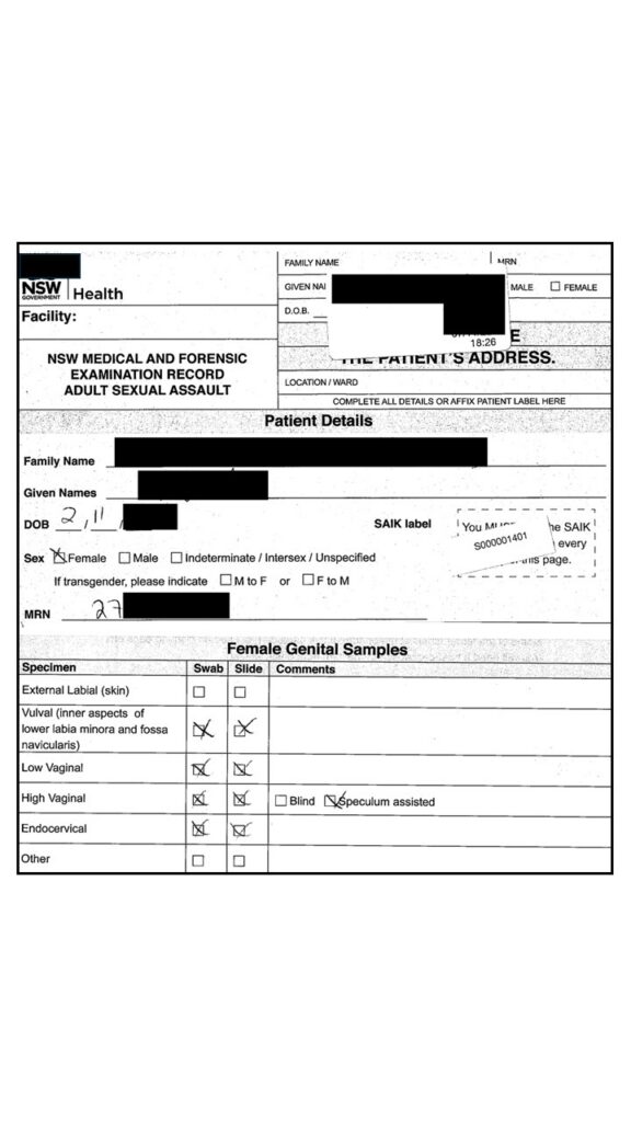
In this case, whilst the laboratory identified semen, the report issued provided no discussion regarding the amount or number of sperm cells detected. The review of the DNA casefile in conjunction with the medical examination records by Ms Roebuck identified that the amount of semen/sperm detected was not in keeping with a full ejaculation of semen into the vagina at the time of the alleged assault. Further, it could not be excluded as a possibility that the semen detected was the result of the previous intercourse between the complainant and her partner.
time since intercourse – semen persistence
Read more scientific detail
TSI evaluation considers both the quantity and distribution of seminal material, the presence and condition of spermatozoa, the anatomical location from which samples were recovered, and the time elapsed between the alleged activity and forensic examination. These factors are interpreted together, rather than in isolation, to assess whether the findings align with the alleged sequence of events or may reasonably be explained by earlier deposition and persistence.
Semen is a complex biological fluid composed of spermatozoa and secretions from the prostate, seminal vesicles and other glands. Following deposition, both the cellular and fluid components undergo biological and mechanical changes. Spermatozoa may degrade, migrate, or be lost over time, while seminal fluid may drain, dilute, or be removed through normal physiological processes such as movement, washing, urination, and menstruation. As a result, the detectability of semen and sperm is not static and varies considerably between individuals and case contexts.
The persistence of spermatozoa within the vaginal tract has been examined extensively in forensic research. Studies demonstrate that intact spermatozoa are most commonly detected within the first 12–24 hours following intercourse, with decreasing recovery rates thereafter, although persistence for several days is possible in some circumstances. The likelihood of detection is influenced by ejaculation, anatomical location, sampling technique, post-event activity, and individual physiological variation. Importantly, the absence of spermatozoa does not exclude prior intercourse, and the presence of spermatozoa does not, in itself, establish when intercourse occurred.
Similarly, the detection of seminal fluid markers, such as acid phosphatase or prostate-specific antigen (PSA), reflects the presence of seminal material but provides limited precision regarding timing. These markers may persist beyond the survival of intact spermatozoa and may be influenced by environmental exposure, substrate, and biological processes. Consequently, laboratory findings indicating semen or sperm must be interpreted cautiously when attempting to relate them to a specific alleged time frame.
In practice, TSI evaluation involves comparing the observed biological findings with what would be expected under different scenarios. For example, a high concentration of intact spermatozoa in anatomically relevant sites may be more consistent with relatively recent intercourse, whereas sparse or degraded spermatozoa, or the detection of seminal fluid without sperm, may be consistent with earlier deposition. These assessments are probabilistic and contextual; they do not provide a precise “time stamp” for sexual activity.
For this reason, TSI evaluation is not determined solely by laboratory detection. It requires consideration of case information, sampling strategy, anatomical context, and the scientific literature on semen persistence and degradation. The purpose is not to assign a definitive time of intercourse, but to assess whether the biological findings support, contradict, or remain neutral in relation to the alleged sequence of events.
In forming opinions regarding timing and persistence, I rely on a substantial body of peer-reviewed forensic research examining sperm survival, seminal fluid persistence, and the effects of post-event activities on biological recovery. This research consistently demonstrates that variability is the norm: recovery outcomes differ between individuals, between anatomical sites, and between cases. As a result, TSI findings must be evaluated cautiously and within the full evidential context, particularly where alternative explanations for the presence of semen or sperm are reasonably available.
Selected research underpinning time-since-intercourse evaluation
- Willott, G.M. (1982). The persistence of seminal constituents in the human vagina. Forensic Science International.
- Davies, A., Wilson, E., & Mitchell, R. (1992). The persistence of seminal material in the female genital tract following intercourse. Medicine, Science and the Law.
- Anderson, S.E., & Langer, S.V. (2005). Time since intercourse studies: spermatozoa and seminal markers in forensic examinations. Forensic Science International.
- Hooft, P., et al. (2009). Persistence of prostate-specific antigen and spermatozoa in vaginal samples following sexual intercourse. Forensic Science International: Genetics.
- Hanson, E., & Ballantyne, J. (2010). A study of the persistence of seminal fluid biomarkers in sexual assault casework. Journal of Forensic Sciences.
- Taylor, D., Biedermann, A., Samie, L., & Hicks, T. (2017). Evaluating forensic biology results in the context of activity level propositions. Forensic Science International: Genetics.
pre ejaculate – Sperm cells without ejaculation
Read more scientific detail
Pre-ejaculatory fluid is a biological secretion released from the penis prior to ejaculation, primarily originating from the bulbourethral (Cowper’s) glands and urethral glands. In forensic examinations, it may be present in the absence of a reported ejaculation and can therefore be relevant in matters where the alleged activity involves sexual contact without ejaculation, interrupted intercourse, or uncertainty regarding what occurred.
Historically, pre-ejaculatory fluid was often assumed not to contain spermatozoa. However, contemporary research demonstrates that this assumption is not scientifically reliable. Studies have shown that sperm may be present in a proportion of pre-ejaculatory samples, even when ejaculation has not occurred immediately beforehand. In one controlled investigation, spermatozoa were detected in pre-ejaculatory fluid from a significant proportion of participants, with some individuals producing samples containing motile sperm. These findings indicate that pre-ejaculatory fluid has the potential to contribute male DNA and, in some cases, spermatozoa to forensic samples.
Scientific studies have reported that up to approximately 41% of men may produce pre-ejaculatory fluid containing spermatozoa under certain conditions. This variability is influenced by individual physiology, prior ejaculation, residual sperm within the urethra, and the timing of sampling. As a result, the presence of sperm within a sample cannot automatically be interpreted as evidence that ejaculation occurred during the alleged event.
From a forensic perspective, the relevance of pre-ejaculatory fluid lies in its capacity to account for the presence of male DNA – and occasionally sperm – in the absence of ejaculation. This is particularly important when evaluating allegations involving digital penetration, genital contact, attempted intercourse, or interrupted sexual activity. The detection of spermatozoa or male DNA may therefore be consistent with a range of activities, not solely full penetrative intercourse with ejaculation.
In activity-level evaluation, this distinction is critical. Laboratory findings can establish that male biological material is present and, in some cases, that spermatozoa have been detected. They cannot determine whether ejaculation occurred, nor can they distinguish between sperm deposited through ejaculation and sperm present within pre-ejaculatory fluid. Consequently, the biological findings must be interpreted within the broader case context, including the alleged sequence of events, the timing of examination, and any alternative explanations for the transfer of biological material.
The persistence and distribution of material associated with pre-ejaculatory fluid may also differ from that observed following ejaculation. Smaller volumes of biological material may be transferred, potentially resulting in lower sperm counts, partial or low-level DNA profiles, or findings limited to external genitalia, clothing, or adjacent surfaces. These outcomes can be consistent with contact or attempted intercourse rather than ejaculation.
For this reason, the presence of spermatozoa or male DNA should not be viewed in isolation when assessing what activity occurred. Instead, the findings must be considered alongside sampling strategy, anatomical location, persistence data, and the scientific literature on semen and pre-ejaculatory fluid. The aim is not to determine a single definitive explanation, but to evaluate whether the biological findings are consistent with the alleged activity, alternative accounts, or both.
In forming opinions on the evidential significance of pre-ejaculatory fluid, I rely on peer-reviewed research examining its composition, the frequency with which spermatozoa are present, and its implications for forensic interpretation. This research demonstrates that pre-ejaculatory fluid can contribute sperm and DNA in a measurable proportion of cases and therefore has direct relevance when evaluating activity-level propositions in sexual assault and related matters.
Selected research underpinning interpretation of pre-ejaculatory fluid:
- Killick, S.R., Leary, C., Trussell, J., & Guthrie, K.A. (2011). Sperm content of pre-ejaculatory fluid. Human Fertility.
2. Zukerman, Z., Weiss, D.B., & Orvieto, R. (2003). Does pre-ejaculatory fluid contain sperm? Journal of Assisted Reproduction and Genetics.
3. Rogerson, C., et al. (2015). The presence and persistence of spermatozoa in low-volume male genital secretions. Forensic Science International: Genetics.
4. Taylor, D., Biedermann, A., Samie, L., & Hicks, T. (2017). Evaluating forensic biology results in the context of activity level propositions. Forensic Science International: Genetics.
How DNA Can Be Transferred During a Medical Examination
Medical examinations are designed to recover biological material, including DNA.
However, the process of sampling itself can introduce complexity, including the potential for DNA to be transferred between locations during collection.
DNA for example can be inadvertently move from the outside of the vagina, to the inside of the vagina by the swab and or speculum.
It is of the utmost importance that stringent DNA anti-contamination procedures are applied when conducting the sampling procedure upon the complainant. The significance in many matters such as this, is less that entirely foreign DNA might be introduced (though that is a consideration and possible), and more that DNA already upon the complainant may be inadvertently transferred during the medical sampling process.
DNA anti contamination procedures include:
- – DNA sterilisation of the environment
- – Documented procedures for cleaning and handling of tools
- – Methodical glove changes at all critical stages
- – Vigilant avoidance of techniques which permit DNA transfer
“In this matter, my expert reporting made mention of numerous factors related to the collection of samples by the medical examiner. The issues raised were ventilated further at trial with the medical examiner, the crown expert and myself all cross examined on issues pertaining to DNA and semen transfer and the use of the speculum. Included in this consideration before the court were numerous key scientific publications”.
transfer from outside to inside vagina – speculum dna transfer – speculum semen transfer
Read more scientific detail
The collection of vaginal swabs during forensic medical examination is a routine and important step in the investigation of alleged sexual assault. These samples are intended to recover biological material that may assist in identifying whether sexual contact occurred and, in some cases, who may have contributed that material. However, the examination process can influence what material is recovered and how it is subsequently interpreted.
During internal examination, swabs are introduced into the vaginal canal either directly or with the assistance of a speculum. In doing so, there is the potential for biological material present on the external genitalia to be transferred internally. This may include epithelial cells, semen, or spermatozoa located on the labia, perineum, or adjacent skin surfaces. Such transfer can occur mechanically during the sampling process, rather than as a result of the alleged activity.
This issue has been examined in controlled forensic research. Experimental studies have demonstrated that DNA and spermatozoa present externally can be introduced into internal vaginal samples during the collection process. In one in-vitro validation study of vaginal sampling methods, external-to-internal transfer of biological material was observed in a substantial proportion of samples. Specifically, approximately 63% of vaginal swabs collected without a speculum showed contamination originating from the external genital region, and 100% of high vaginal swabs collected using a speculum demonstrated transfer of DNA from external sites. These findings highlight the potential for sampling-related transfer to occur even under controlled conditions.
More recent work from the United Kingdom has reinforced these findings in contemporary forensic medical settings. Validation studies conducted within Sexual Assault Referral Centres (SARCs) have examined how vaginal and ano-genital samples are collected in practice and the pathways through which biological material may be recovered or redistributed during examination. This body of work recognises sampling as both a recovery process and a potential transfer mechanism, and emphasises the importance of contamination control, sampling order, and examination technique. The findings support the conclusion that biological material detected in internal vaginal samples may, in some circumstances, reflect redistribution from external genital sites during the examination process itself.
The relevance of this phenomenon is grounded in a fundamental forensic principle: biological material can be transferred through contact. While this principle is often discussed in relation to alleged activity, it also applies to the forensic examination process itself. When swabs are collected, the pathway created by instruments, swabs, and handling may facilitate movement of material from one anatomical site to another.
From an interpretive perspective, this means that the detection of DNA or spermatozoa within internal vaginal samples does not necessarily establish that those materials were deposited internally during the alleged event. In some circumstances, the findings may instead reflect redistribution of material that was present externally at the time of examination. This is particularly relevant where semen or male DNA is detected internally but where questions arise regarding the mechanism, timing, or location of deposition.
The distinction between primary deposition and sampling-related transfer is an activity-level question. Laboratory testing can identify the presence of DNA or spermatozoa within a swab, but it cannot determine whether those materials arrived at that location during the alleged activity or during the medical examination. The evaluation therefore requires consideration of examination notes, sampling technique, anatomical context, and the broader case circumstances.
In practice, this does not diminish the value of forensic medical sampling. Rather, it underscores the importance of understanding how samples are collected and how biological material may move between sites. Where internal findings are relied upon as evidence of penetration or ejaculation, the possibility of transfer during examination must be considered as part of a balanced forensic evaluation.
In forming opinions on the evidential significance of internal vaginal findings, I rely on peer-reviewed research examining sampling techniques, transfer mechanisms, and the performance of forensic medical examinations. These studies demonstrate that biological material can be redistributed during swab collection and that this has direct implications for how internal findings are interpreted in relation to alleged activity.
Selected research underpinning transfer during vaginal sampling:
- Loeve, A.J., Bilo, R.A.C., Emirdag, E., et al. (2013). In vitro validation of vaginal sampling in rape victims: the problem of Locard’s principle. Forensic Science, Medicine and Pathology, 9:154–162.
- Recent UK validation studies conducted within Sexual Assault Referral Centres examining DNA recovery pathways, sampling technique, and contamination risk during ano-genital forensic examination.
- Faculty of Forensic & Legal Medicine (UK). Guidance on the order and practice of ano-genital sampling in forensic medical examinations, addressing contamination control and transfer risk in sexual offence investigations.
Laboratory Analysis of Vaginal Swabs
While the medical examiner collects the vaginal swabs and prepares microscope slides, it is ultimately the forensic laboratory that conducts the detailed examination and testing of those samples.
Vaginal swab DNA evidence is commonly relied upon in sexual assault cases because these samples may seek to identify semen, sperm cells, or male DNA profiles.
The strategic approach to testing vaginal swabs is complex, with several possible pathways.
The pathway applied can effect the results.
In general, laboratories first screen swabs for indicators of seminal fluid, followed by confirmatory examination where results suggest semen may be present. This typically includes microscopic assessment for spermatozoa and, where appropriate, further biochemical or DNA testing.
In this case, preliminary screening indicated the possible presence of seminal fluid on the low vaginal swab and the vulval swab. No spermatozoa were identified on the low vaginal swab, and only a single sperm cell was observed on the vulval swab. The identification of even a single sperm head is generally accepted as confirmation that semen is present on that swab.
Understanding the testing performed, and the results obtained in their entirety, is critical when evaluating what these findings do – and do not – say about the allegation. The location of the findings, the quantity of sperm observed, and the relationship between screening results, microscopy and DNA testing all inform whether the swab results meaningfully support the alleged events.
Laboratory reports summarise results.
They do not evaluate how those results fit the allegation or background circumstances.
That interpretation occurs later — often for the first time at trial.
Clothing Handling and the Potential for DNA Transfer Before Police Seizure
Appropriate handling, packaging and storage of items for DNA testing is critical.
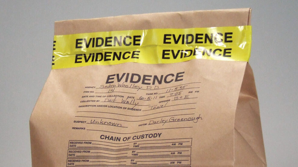
Upon reporting the matter to police, the complainant made it known that she was not wearing the same underpants and clothing that she was wearing at the time of the alleged incident.
The complainant stored, handled and packaged those underpants at her home, before delivering them to police.
This handling and potential for DNA transfer complicated the case.
Further statements of the complainant were reviewed which provided finer detail from Ms Hood. After the alleged incident, at around 2 to 3am, the complainant remained in the same clothing, drifting back to sleep on the couch until sometime around 9 or 10am the same day. Upon waking, she urinated and exchanged numerous text messages with a friend about the alleged incident. She then removed all of her clothing (including the underpants) and left them in a pile on the living room floor. Without showering, she dressed in clean clothing and attended the police station. After attending the police station, she was medically examined and then returned to her home to collect the underpants. The complainant delivered the underpants to police the following day at 10am in a Coles plastic shopping bag.
This information is crucial when evaluating the DNA results, as it is known that DNA can transfer between garments, be relocated upon an item; and even be transferred from one area of the item to another when packaged in police evidence bags.
Transfer during packaging – poor handling – contamination with and without gloves
Read more scientific detail
Forensic exhibit packaging is designed to preserve evidence, prevent contamination, and maintain the integrity of items from the point of collection through to laboratory examination. Packaging is often perceived as a protective barrier — a step that “locks in” the evidential value of an item. In practice, however, packaging is not a static environment. Biological material can move, be redistributed, or be lost during handling, transport, and storage.
DNA is not fixed to a surface once deposited. Cellular material can detach, transfer, and relocate within an exhibit package through routine movement. Friction, vibration, handling, folding, and contact between items or between an item and its packaging material can all contribute to redistribution. This means that DNA detected on a particular area of an exhibit may not necessarily reflect the original site of deposition.
Experimental research has demonstrated that DNA relocation within packaging is a measurable and recurring phenomenon. Studies examining forensic packaging conditions have shown that DNA can move from one area of an exhibit to another during storage and transport, and that this process may result in both loss of DNA from the original site and deposition in new locations. In controlled experiments, relocation of DNA within exhibit packaging was observed in approximately 39% of packaged items, highlighting that such movement is not rare and may occur under routine handling conditions.
More recent work has further demonstrated that transfer can also occur between items stored within the same evidence package. Where multiple exhibits are packaged together – for example, clothing items, bedding, or personal effects – biological material may move between substrates through contact and motion. This has implications for the interpretation of DNA findings, particularly where the location of DNA on an item is relied upon as evidence of direct contact or activity.
From an interpretive perspective, this means that the presence of DNA on a specific region of an exhibit does not automatically establish how or when it was deposited. Redistribution during packaging and storage may alter the original pattern of biological material, potentially creating findings that appear to reflect contact with that location when, in fact, the DNA may have originated elsewhere on the item — or from another item within the same package.
The integrity of packaging is therefore directly relevant to activity-level evaluation. Laboratory testing can determine that DNA is present and may identify its source, but it cannot determine whether that DNA remained in its original location from the time of deposition. Consideration must be given to how the exhibit was packaged, whether items were stored together, how frequently they were handled, and the duration and conditions of storage.
This issue is particularly important in matters where the location of DNA on an item is used to support an allegation – for example, DNA detected on a specific region of clothing, bedding, or an object said to have been handled in a particular way. Redistribution within packaging may provide an alternative mechanism for how DNA came to be present at that location.
For this reason, the evaluation of DNA findings requires examination not only of laboratory results but also of packaging records, continuity documentation, and handling history. The way in which exhibits were packaged, transported, and stored can materially influence what biological material is ultimately detected and where it is found.
In forming opinions on the evidential significance of DNA detected on exhibits, I rely on peer-reviewed research examining transfer mechanisms, persistence, and the behaviour of DNA during storage and handling. This research demonstrates that relocation within packaging can occur under routine conditions and that such movement must be considered when interpreting DNA findings in the context of alleged activity.
Selected research underpinning DNA relocation within exhibit packaging
-
Goray, M., van Oorschot, R.A.H., & Mitchell, J.R. (2012). DNA transfer within forensic exhibit packaging: Potential for DNA loss and relocation. Forensic Science International: Genetics, 6(2), 158–166.
-
Lee, Y.S., & Syn, C.K.-C. (2025). DNA transfer between items within an evidence package. Genes, 16(8), 894.
Sampling Strategy: Where and Why Items Are Tested
The method and location of sampling can significantly affect the results.
The examination of any item should follow what is forensically termed a sampling strategy.
In simple terms, this means that a deliberate approach is developed before testing begins.
This strategy should consider what results might reasonably be expected, what is being alleged, how the item was handled, and what questions the examination is intended to answer.
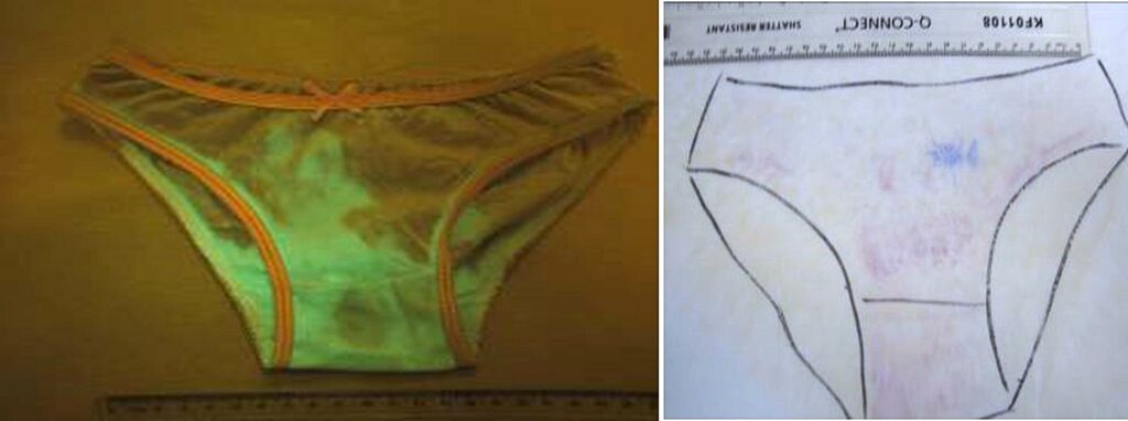
In the case of these underpants, the sampling strategy would have ideally been informed by how the complainant reported handling the garment, including when it was worn, removed, and stored. That context can be critical in determining where relevant biological material might reasonably be located.
In this matter, the sampling notes and cross examination of the sampling officer, indicated that the sampling approach was not formulated with this information in mind. This is not uncommon. In practice, laboratory sampling is often guided by standard workflows designed to maximise the chance of recovering DNA, rather than by a tailored strategy focused on the evidential questions raised by the allegation. No semen was detected on the underpants, and the hips and gusset of the underwear were sampled in an attempt to identify DNA from individuals whom may have contacted the clothing.
Hip Area Sampling
The hips of underwear are often sampled due to the high chance of success in raising a DNA profile.
Hips are routinely contacted during adjustment.
A female wearer will contact the hips multiple times during each toileting event.
The sampling officer’s statement and notes did not articulate whether the sampling of the hips was of the outside or inside hips; limiting the ability to interpret the DNA findings.
A DNA profile matching Mr Smith was obtained from the sample taken from the hips. Importantly, it should be recalled that there was extensive social contact between the individuals, with Mr Smith placing his arm around her waist.
Gusset Area Sampling
In this case, the “inside gusset” of the underwear was sampled for DNA, as was the “upper front”.
The sampling officer’s statement and notes failed to identify whether the upper front was the inside or outside.
Inside or outside of underwear can be critically relevant.
Further, this sample was combined with the gusset sample for DNA testing. This means that the DNA result from this sample may be from the “upper front outside” or “upper front inside” or “inside gusset” or indeed any unknowable combination.
For these reasons, the sample being known in evidence as the “gusset sample” is at best imprecise and at worst misleading.
The DNA profile obtained from the “gusset” matched that of Ms Hood. There was no evidence that Mr Smith had contributed to the DNA profile.
“My reporting in this matter made extensive reference to the sampling strategy applied to the underpants. There was significance in the handling of the underpants prior to arrival with police. Specifically, how that handling may impact upon the propensity for DNA present upon the other clothing, couch and outer surfaces of the underwear (including the hips) to transfer inadvertently to the gusset of the underpants. These issues were canvassed extensively at trial with the police sampling officer, the crown expert and myself, all cross examined. Further, the crown expert and myself were questioned on DNA transfer during toileting and semen drainage from the vagina, and semen transfer from underwear to the vulva whilst worn”.

Sample targeting – Sampling strategy
Read more scientific detail
The sampling strategy adopted during forensic examination is a critical component of DNA evidence generation and interpretation. In this matter, the high and low vaginal swabs were combined prior to testing. This approach affects how the biological material is represented in subsequent laboratory results and has implications for the interpretation and evidential value of any DNA findings.
Determining an appropriate sampling strategy requires consideration of multiple factors, including the allegation, the anatomical location of interest, the expected distribution of biological material, the time since the alleged activity, and the potential for transfer or degradation. In practice, however, laboratory workflows are often oriented toward maximising the likelihood of obtaining a DNA profile. While this is an important operational objective, it is distinct from the question of evidential value – that is, what the resulting DNA findings can meaningfully say about the alleged events.
Once swabs from different anatomical locations are combined, the ability to attribute any resulting DNA profile to a specific site is lost. If a DNA profile is subsequently obtained, it is no longer possible to determine whether it originated from the high vaginal region, the low vaginal region, or a combination of both. In certain case contexts, this distinction may be scientifically and contextually important, as the distribution of biological material within the vaginal canal can vary depending on time since intercourse, movement, drainage, and other post-event factors.
In this matter, no DNA profile was ultimately obtained from the combined swabs, and the impact of the sampling strategy is therefore limited. However, had a profile been generated, its anatomical origin would have remained uncertain. This may have influenced the interpretation of the findings, particularly if questions arose regarding persistence, timing, or the mechanism of deposition.
Laboratory sampling strategies are not described in detail within forensic reports. Nonetheless, the case file, examination notes, and laboratory documentation can provide insight into how samples were collected, processed, and prioritised. In reviewing this material, I considered whether the approach taken was appropriate in the context of the case, and whether alternative sampling or testing strategies may have been available. In some circumstances, targeted examination of individual swabs, or the use of complementary recovery techniques, may assist in identifying the presence – or absence – of biological material in specific locations and may provide information of potential evidential value, including exculpatory findings.
A range of established recovery methods are available in forensic biology, including site-specific swabbing, staged sampling, and adhesive tapelift techniques for recovering cellular material from skin or substrates. The selection of method influences both the likelihood of recovering DNA and the interpretive value of the results. For this reason, sampling strategy is not merely a technical step in the laboratory process; it is integral to how forensic findings are generated, understood, and ultimately evaluated in the context of the case.
In forming opinions on sampling and testing strategy, I rely on a substantial body of peer-reviewed research addressing evidence recovery methods, anatomical sampling, and the performance of swabbing and tapelift techniques in forensic casework. These studies demonstrate that recovery outcomes and interpretive value are influenced by sampling location, method selection, and handling practices, and that different strategies may produce materially different evidential outcomes.
1. Sewell, J., & Quinones, I. (2014). Collection of biological evidence in sexual assault investigations. Journal of Forensic Nursing.
2. Verdon, T.J., Mitchell, R.J., & van Oorschot, R.A.H. (2014). The influence of sample type and DNA extraction method on the detection of DNA in mixed biological samples. Forensic Science International: Genetics.
3. Foster, E.A., et al. (2013). Evaluation of swabbing techniques for the recovery of DNA from skin surfaces. Forensic Science International: Genetics.
4. Daly, D.J., Murphy, C., & McDermott, S.D. (2012). The transfer of touch DNA and the effectiveness of recovery techniques. Forensic Science International: Genetics.
5. Fieldhouse, S., & Gwinnett, C. (2012). The use of tapelifts in the recovery of DNA evidence from surfaces. Journal of Forensic Identification.
6. Rutty, G.N. (2001). An investigation into the effectiveness of swabbing and tapelift techniques in forensic sampling. Forensic Science International.
What a LAB DNA Report Actually Includes
In NSW an expert certificate must be served for the DNA to be relied upon in evidence. In other states of Australia the comparable reporting is known by other titles such as expert report in Victoria and Queensland.
All states report similarly in the respect that reporting is commonly limited to source (who).
Reporting is typically very brief and will not evidence all testing or methods applied.
Regardless of the title, the principle is the same in that the named expert upon the report may be called to give oral evidence in support of the report, should the matter proceed to trial. The remainder of the report is made of appendixes which cover related general scientific constructs such as DNA transfer, however the expert has not applied those considerations to the background information in the matter.
Though the expert report does not contain matter specific opinions or broader scientific considerations of DNA transfer and research papers, one should expect that these issues will be live and opined upon at trial by the author of the report.
Of note, whilst the expert report seems quite simple on the face of it, there is significant work conducted by the lab leading up to this final report. This underpinning material is not issued with the expert report, however it is obtainable of the laboratory, generally by subpoena.
In this matter, whilst the DNA report was only two pages long, the DNA casefile material obtained for the independent review encompassed some 220 pages, which includes the electropherograms, contemporaneous examination records, laboratory analysis results, probabilistic genotyping outputs and haplotype database counts
What The lab DNA Report Does Not Establish
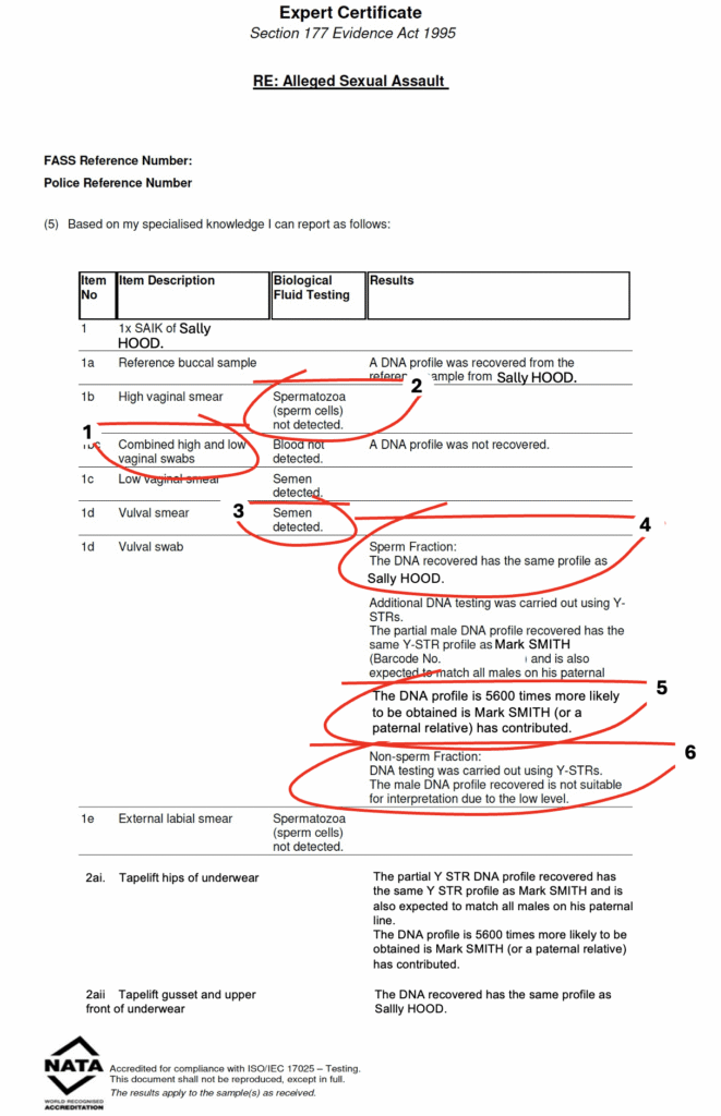
‘A common misunderstanding is to read DNA reports as absolute. In my experience, most non forensic biologists would take the below expert report to mean that there is sperm cells and DNA within the vagina, and that the sperm cells are highly likely to have originated from Mark Smith – The below report does not establish those things’
Why DNA results can look stronger than they really are
DNA reports are written to describe laboratory findings.
They do not:
- – Decide whether an allegation occurred
- – Determine how DNA was deposited
- – Assess competing explanations
- – Evaluate transfer scenarios
- – Apply the broader case background
This is why DNA evidence that appears strong in a report can later weaken significantly when examined in context.
→ Read: When DNA statistics are wrong
→ Read: How courts assess whether DNA should be heard at trial
What the DNA report Actually means
The numbered points below explain the numbered points on the DNA expert certificate above. Importantly, the true understanding cannot be obtained from the face of expert report itself, rather through evaluating the DNA casefile material in this specific matter in consideration of the relevant scientific literature. That fuller scientific methodology to each specific point is detailed further in the read more scientific detail at each numbered section below.
‘
The medical examiner obtained a swab from the high vagina and a swab from the low vagina.
These two swabs were combined by the laboratory and tested for DNA.
There is no evidence that Mr Smith’s DNA is inside the vagina.
Read more Scientific detail
The sampling strategy adopted during forensic examination is a critical component of DNA evidence generation and interpretation. In this matter, the high and low vaginal swabs were combined prior to testing. This approach affects how the biological material is represented in subsequent laboratory results and has implications for the interpretation and evidential value of any DNA findings.
Determining an appropriate sampling strategy requires consideration of multiple factors, including the allegation, the anatomical location of interest, the expected distribution of biological material, the time since the alleged activity, and the potential for transfer or degradation. In practice, however, laboratory workflows are often oriented toward maximising the likelihood of obtaining a DNA profile. While this is an important operational objective, it is distinct from the question of evidential value – that is, what the resulting DNA findings can meaningfully say about the alleged events.
Once swabs from different anatomical locations are combined, the ability to attribute any resulting DNA profile to a specific site is lost. If a DNA profile is subsequently obtained, it is no longer possible to determine whether it originated from the high vaginal region, the low vaginal region, or a combination of both. In certain case contexts, this distinction may be scientifically and contextually important, as the distribution of biological material within the vaginal canal can vary depending on time since intercourse, movement, drainage, and other post-event factors.
In this matter, no DNA profile was ultimately obtained from the combined swabs, and the impact of the sampling strategy is therefore limited. However, had a profile been generated, its anatomical origin would have remained uncertain. This may have influenced the interpretation of the findings, particularly if questions arose regarding persistence, timing, or the mechanism of deposition.
Laboratory sampling strategies are not described in detail within forensic reports. Nonetheless, the case file, examination notes, and laboratory documentation can provide insight into how samples were collected, processed, and prioritised. In reviewing this material, I considered whether the approach taken was appropriate in the context of the case, and whether alternative sampling or testing strategies may have been available. In some circumstances, targeted examination of individual swabs, or the use of complementary recovery techniques, may assist in identifying the presence – or absence – of biological material in specific locations and may provide information of potential evidential value, including exculpatory findings.
A range of established recovery methods are available in forensic biology, including site-specific swabbing, staged sampling, and adhesive tapelift techniques for recovering cellular material from skin or substrates. The selection of method influences both the likelihood of recovering DNA and the interpretive value of the results. For this reason, sampling strategy is not merely a technical step in the laboratory process; it is integral to how forensic findings are generated, understood, and ultimately evaluated in the context of the case.
In forming opinions on sampling and testing strategy, I rely on a substantial body of peer-reviewed research addressing evidence recovery methods, anatomical sampling, and the performance of swabbing and tapelift techniques in forensic casework. These studies demonstrate that recovery outcomes and interpretive value are influenced by sampling location, method selection, and handling practices, and that different strategies may produce materially different evidential outcomes.
1. Sewell, J., & Quinones, I. (2014). Collection of biological evidence in sexual assault investigations. Journal of Forensic Nursing.
2. Verdon, T.J., Mitchell, R.J., & van Oorschot, R.A.H. (2014). The influence of sample type and DNA extraction method on the detection of DNA in mixed biological samples. Forensic Science International: Genetics.
3. Foster, E.A., et al. (2013). Evaluation of swabbing techniques for the recovery of DNA from skin surfaces. Forensic Science International: Genetics.
4. Daly, D.J., Murphy, C., & McDermott, S.D. (2012). The transfer of touch DNA and the effectiveness of recovery techniques. Forensic Science International: Genetics.
5. Fieldhouse, S., & Gwinnett, C. (2012). The use of tapelifts in the recovery of DNA evidence from surfaces. Journal of Forensic Identification.
6. Rutty, G.N. (2001). An investigation into the effectiveness of swabbing and tapelift techniques in forensic sampling. Forensic Science International.
Microscopy was conducted and no sperm cells were detected upon the high vagina swab.
A full healthy male ejaculate can contain millions of sperm heads and would be expected to deposit observable sperm at the high vagina.
As the swabs were taken approximately 13 hours after the alleged sexual intercourse (assuming ejaculation into the vagina occurred), there would be a reasonable expectation for observed sperm cells at the high vagina.
As the swabs were taken 24 to 36 hours after the complainant had sexual intercourse with her partner (assuming ejaculation into the vagina occurred), we would not necessarily expect sperm cells at the high vagina.
Read more Scientific detai
No spermatozoa were detected on the high vaginal swab in this matter. In those circumstances, semen cannot be confirmed at that site on the basis of the laboratory findings. The absence of sperm cells is a scientifically relevant observation, but it does not, in isolation, establish that ejaculation did not occur.
The persistence of spermatozoa within the vaginal environment is known to be time-dependent and highly variable. Following ejaculation, sperm cells may be progressively lost, degraded, or redistributed through a combination of biological and mechanical processes. These include natural drainage, vaginal physiology, microbial activity, enzymatic degradation, and the effects of post-event activities such as washing, urination, menstruation, or further consensual sexual activity. The timing of sampling relative to the alleged event is therefore critical when interpreting the absence of spermatozoa.
Time-since-intercourse research demonstrates that sperm recovery rates decline over time and that detection is influenced by multiple variables, including the volume of ejaculate, condom use, anatomical differences, movement and posture, and the collection method used during examination. Even within relatively short timeframes, spermatozoa may not be recovered from all vaginal sampling sites, and distribution within the vaginal canal is not uniform. The absence of sperm on a high vaginal swab must therefore be considered holistically alongside findings from other sampling locations, laboratory results, and the broader case circumstances.
Importantly, the absence of spermatozoa does not preclude the presence of seminal fluid or male DNA. Sperm cells may degrade or be absent while other components of semen — including proteins, enzymes, or non-sperm cellular material — persist and may still be detectable through biochemical or DNA testing. For this reason, interpretation must consider the full range of laboratory findings rather than relying on microscopy alone.
In forming opinions on the absence of spermatozoa and its significance, reliance is placed on a substantial body of peer-reviewed research examining sperm persistence, time-since-intercourse dynamics, vaginal biology, and the recovery of semen and DNA following sexual activity. These studies consistently demonstrate that sperm detection is influenced by time, environment, and post-event factors, and that non-detection cannot be interpreted in isolation. This literature provides the scientific framework for evaluating the evidential significance of the findings in this matter.
1 Willott, G.M. (1989). An examination of the persistence of spermatozoa in the vagina. Forensic Science International.
2 Davies, A., & Wilson, E.M. (1974). The persistence of seminal constituents in the human vagina. Forensic Science.
3 Hanson, E.K., & Ballantyne, J. (2010). Time since intercourse: the challenge of sperm persistence and recovery in sexual assault casework. Forensic Science International: Genetics.
4 Mayntz-Press, K.A., & Morling, N. (2015). Forensic investigation of sexual assault – time since intercourse and biological evidence. Forensic Science International: Genetics.
5 Girardet, R., et al. (2009). Collection and persistence of biological evidence following sexual assault. Journal of Forensic Sciences.
6 Anderson, S., Howard, B., Hobbs, G.R., & Bishop, C.P. (2006). A method for determining the time since intercourse from biological samples. Forensic Science International.
The low vagina semen result is a presumptive test only, meaning no sperm cells at the low vagina.
Within the DNA casefile, microscope work was conducted and 1 sperm cell was identified on the vulval swab.
The complainant could not confirm how long she wore the underpants following the 24 to 36 hour intercourse with her partner.
The external vaginal 1 sperm head is considerably complicated by the persistence and transfer issues related to the worn underpants.
Read more Scientific detail
Spermatozoa are microscopic reproductive cells produced in the testes. Only a small portion of semen is made up of these cells; the majority is a complex biological fluid containing proteins, enzymes, and other biochemical components contributed primarily by the prostate gland, seminal vesicles, and other accessory glands.
Importantly, semen does not always contain sperm cells. Conditions such as vasectomy, certain medical treatments, age, or naturally low sperm counts (including azoospermia) can result in seminal fluid being present in the absence of detectable spermatozoa. A study by Soares-Vieira et al. (2007) analysed the DNA content of semen from 105 males, determining that the concentration of DNA present varied substantially. Therefore the visualisation of sperm cells within the sample is crucial to the interpretation of the forensic DNA results.
Laboratory testing often targets specific biochemical markers that are characteristic of seminal fluid. Key components include enzymes and proteins such as acid phosphatase, prostate-specific antigen (PSA), and semenogelin.
Acid phosphatase is an enzyme produced in high concentrations by the prostate gland. Presumptive acid phosphatase testing is commonly used as an initial screening method because elevated enzyme activity may indicate the presence of seminal fluid. Early work Early work by Kind et al (1954) demonstrated that the acid phosphatase test has practical detection thresholds and may fail to identify very dilute or degraded seminal material. The limitations of this test are also extensively reported in the literature; and an intimate understanding of the methodology applied within the specific laboratory assists in identifying the potential for false positive reactions in the specific matter.
Prostate-specific antigen (PSA), also known as p30, is a protein produced by prostatic epithelial cells and is sometimes used as a more specific marker for seminal fluid. PSA testing can detect semen even where spermatozoa are absent, but it too is not entirely unique to semen and may occasionally be identified in other bodily fluids at low concentrations.
Semenogelin, the target protein of the RSID Semen test is a major structural protein derived from the seminal vesicles and forms the coagulated matrix of freshly ejaculated semen. Its presence can assist in confirming seminal material and in understanding the biochemical processes involved in semen liquefaction and degradation over time.
Understanding the biological function and origin of these enzymes and proteins is central to forensic interpretation. Laboratory tests do not directly answer whether ejaculation occurred, when it occurred, or the circumstances in which biological material was deposited. Rather, they detect markers consistent with seminal fluid, each with its own specificity, sensitivity, and potential for cross-reactivity. The interpretation of results therefore requires careful consideration of the underlying biochemistry, the limitations of each test, and the broader evidential context in which the findings are being applied
.
DNA sample is separated into to portions; referred to as a sperm fraction and a non-sperm fraction.
These terms can be misleading as the sperm fraction does not necessarily contain sperm.
Processing the sample in this manner is intended to assist with determining whether the DNA profile is reasonably attributable to the 1 sperm head observed.
In this matter, the processing of the sperm fraction and non sperm fraction failed, as female DNA was carried into the sperm fraction.
The 1 sperm cell cannot be said to have originated from Mark Smith.
Read more Scientific detail
The presence of the terms sperm fraction and non-sperm fraction in a forensic DNA report indicates that a process known as differential extraction was used during laboratory testing. This is important, as it provides insight into how the sample was handled and how the resulting DNA profiles were generated.
Differential extraction is a laboratory technique designed to separate sperm cells from other cellular material within a biological sample, most commonly in the examination of sexual assault evidence. The aim is to preferentially recover male DNA from spermatozoa while isolating DNA from epithelial and other non-sperm cells into a separate fraction.
The process is carried out in stages. Initially, non-sperm cells — such as skin or vaginal epithelial cells — are lysed using detergents and enzymes, releasing their DNA into solution. This material is collected as the non-sperm fraction. The remaining cellular material is then treated with additional reagents, including the reducing agent dithiothreitol (DTT), which disrupts the structural integrity of sperm heads and allows DNA from spermatozoa to be released and collected separately as the sperm fraction. DTT is required because spermatozoa are resistant to standard lysis; the DNA within the sperm nucleus is tightly compacted by protamines and stabilised by extensive disulfide cross-linking within sperm head proteins.
However, this method is not perfectly efficient. Carryover of DNA from non-sperm cells into the sperm fraction is well recognised. In this matter, DNA attributable to a female contributor is detected within the sperm fraction, despite not originating from spermatozoa themselves. This observation is consistent with the known limitations of differential extraction and must be considered in interpretation.
For this reason, the presence of DNA within the sperm fraction must be approached cautiously. It does not automatically mean that the DNA originated from sperm, nor does it, in isolation, establish direct sexual contact. DNA detected in this fraction may arise from other biological material, including epithelial cells introduced through prior or indirect contact, or through transfer during handling and processing. Scientific interpretation is therefore required to understand what the findings do — and do not — indicate.
Interpretation in this matter is further complicated by the case file recording the identification of only a single spermatozoon on the vulval swab. In such circumstances, careful consideration must be given to whether the DNA profile observed in the electropherogram represents the expected outcome if the DNA were derived from sperm, as opposed to another cellular source. This type of evaluation requires detailed knowledge of the extraction process, the behaviour of mixed cellular material during laboratory processing, and the way DNA from different sources is represented in resulting profiles.
In reviewing the evidence and forming my opinions, I rely on an extensive body of peer-reviewed scientific literature addressing differential extraction, sperm cell lysis, DNA recovery from mixed biological samples, and the interpretation of sperm and non-sperm fractions in forensic casework. Foundational studies describing the development and performance of differential extraction methods, research examining the effects of carryover between fractions, and contemporary work on DNA interpretation in complex mixtures collectively inform the evaluation of laboratory findings. These sources provide the scientific framework within which the results in this matter are assessed and interpreted.
Differential extraction techniques, their efficiency, and their limitations in mixed biological samples have been extensively described in the forensic literature (Gill et al.; Yoshida et al.; Horsman et al.; Verdon et al.; Norris et al.; Garvin et al.; Castro et al.).
1 Gill, P., Jeffreys, A.J., & Werrett, D.J. (1985). Forensic application of DNA ‘fingerprints’. Nature, 318, 577–579.
2 Yoshida, K., Sekiguchi, K., & Mizuno, N. (1995). A simple method for differential extraction of sperm DNA from mixed biological stains. Journal of Forensic Sciences.
3 Horsman, K.M., Barker, S.L., Ferrance, J.P., Forrest, K.A., Koen, K.A., & Landers, J.P. (2006). Separation of sperm and epithelial cells in a microfabricated device: potential application to forensic analysis of sexual assault evidence. Analytical Chemistry, 78, 3122–3128.
4 Verdon, T.J., Mitchell, R.J., van Oorschot, R.A.H. (2014). The influence of sample type and DNA extraction method on the detection of male DNA in mixed samples. Forensic Science International: Genetics.
5 Norris, J.V., Manning, K., & Linke, S.J. (2007). Evaluation of differential extraction methods for forensic DNA analysis of sexual assault samples. Journal of Forensic Sciences.
6 Garvin, A.M., Bottinelli, M., Gola, M., Conti, A., & Soldati, G. (2009). DNA preparation from sexual assault cases by selective degradation of contaminating DNA from the victim. Forensic Science International: Genetics
7 Castro, A., Alonso, A., & Martín, P. (2014). Challenges in differential extraction and interpretation of sperm and epithelial fractions in forensic casework. Forensic Science International: Genetics.
8 Green, R.L., & Sambrook, J. (2012). Molecular cloning: laboratory approaches to DNA extraction and protein denaturation relevant to sperm lysis.
This number is what is referred to as a likelihood ratio.
In forensic DNA testing, a likelihood ratio is a scientific expression of how much more probable the observed DNA results are under one proposition compared with an alternative. In an inclusionary sense, likelihood ratios range from 1 to 100 billion, meaning that this number is towards the lower end of the scale.
Read more Scientific detail
This number is what is known as a likelihood ratio. In forensic DNA testing, a likelihood ratio is a scientific expression of how much more probable the observed DNA results are under one proposition compared with an alternative. Most commonly, the comparison is between the proposition that the DNA originated from a named individual and the proposition that it originated from an unknown, unrelated person. It is a measure of the strength of the scientific findings; it is not a statement about guilt, and it does not explain how or when the DNA was deposited.
In this matter, the likelihood ratio evaluates two competing propositions: that the DNA originated from the accused (or one of his paternal relatives), or that it originated from an unknown, unrelated individual. It does not address the mechanism of transfer, the timing of deposition, or the activity that led to the DNA being present. It speaks only to the relative probability of the DNA findings under the competing propositions about source.
A likelihood ratio of 5,600 means that the DNA results are 5,600 times more probable if the DNA came from the named individual (or a paternal relative) than if they came from someone unrelated. In scientific terms, this is typically described as providing strong support for the proposition that the DNA originated from that paternal lineage, within the limits of the assumptions and propositions being tested.
When assessing the robustness of a likelihood ratio, several factors must be considered. These include the quality and completeness of the DNA profile, the number of contributors present in the sample, the allele frequency databases used, and the population groups selected for statistical evaluation. Population genetics is central to likelihood ratio calculations: the rarer a genetic profile is within a relevant population, the stronger the statistical support that may be attributed to a match. The choice of database and the representativeness of the reference populations therefore directly influence the resulting likelihood ratio.
The type of DNA testing used is also critical. In autosomal DNA analysis, likelihood ratios are often generated using probabilistic genotyping software, such as STRmix, which applies complex statistical models to interpret mixed DNA profiles. These systems incorporate allele frequency databases, account for stochastic effects such as allele drop-out and drop-in, and evaluate multiple competing contributor combinations. While such methods are scientifically validated and widely used, they rely on modelling assumptions and population data. The possibility of false inclusions – where an individual may appear to be supported by the DNA evidence due to statistical modelling, mixture complexity, or shared alleles within a population – must therefore always be considered alongside the reported likelihood ratio.
In contrast, Y-STR haplotype evidence is interpreted differently from autosomal DNA. Because Y-STR profiles are inherited along the paternal line and shared among male relatives, statistical evaluation is based on haplotype frequency rather than independent allele combinations. Likelihood ratios derived from Y-STR testing therefore reflect the frequency of that paternal lineage within relevant populations, rather than the probability of a unique individual profile. As a result, Y-STR results cannot distinguish between fathers, sons, brothers, or other paternal relatives, all of whom may be potential sources of the DNA.
Accordingly, a likelihood ratio based on Y-STR findings must be interpreted within this framework. It can indicate that the DNA is consistent with a particular paternal lineage and that such a lineage may be relatively uncommon in the reference population, but it cannot identify a specific individual to the exclusion of all other males within that lineage, nor can it determine the circumstances in which the DNA was deposited.
In reviewing the likelihood ratio evidence and forming my opinions, I rely on a substantial body of peer-reviewed scientific research and validation data relating to population genetics, allele frequency databases, probabilistic genotyping, and the statistical interpretation of DNA mixtures. This includes studies examining the performance and limitations of likelihood ratio approaches, the operation and validation of software such as STRmix, the risk of false inclusions in complex mixtures, and the interpretation of Y-STR haplotype frequencies using large reference datasets. These sources provide the scientific and statistical foundation for evaluating the strength, reliability and limitations of likelihood ratio evidence in the context of the specific circumstances of the case.
1. Evett, I.W., Gill, P.D., Jackson, G., Whitaker, J., & Champod, C. (2002). Interpreting DNA evidence: a review. International Statistical Review, 70(3), 473–495.
2. Buckleton, J., Triggs, C.M., & Walsh, S.J. (2005). Forensic DNA Evidence Interpretation. CRC Press.
3. Gill, P., Brenner, C., Buckleton, J., et al. (2006). DNA commission of the International Society of Forensic Genetics: recommendations on the interpretation of mixtures. Forensic Science International, 160, 90–101.
4. Bright, J.A., Taylor, D., Curran, J.M., & Buckleton, J. (2016). Developing allelic and stutter peak height models for a continuous method of DNA interpretation. Forensic Science International: Genetics, 21, 13–22.
5. Taylor, D., Bright, J.A., & Buckleton, J. (2013). The interpretation of single source and mixed DNA profiles. Forensic Science International: Genetics, 7, 516–528.
6. Bleka, Ø., Storvik, G., Gill, P., & Egeland, T. (2016). EuroForMix: an open source software based on a continuous model to evaluate STR DNA profiles from a mixture. Forensic Science International: Genetics, 21, 35–44.
7. Taylor, D., Bright, J.A., Buckleton, J., & Curran, J.M. (2013). An illustration of the use of STRmix™ for DNA mixture interpretation. Forensic Science International: Genetics, 7(2), 226–233.
8. Willuweit, S., & Roewer, L. (2015). The new Y Chromosome Haplotype Reference Database (YHRD): an extended online resource for Y-STR haplotypes. Forensic Science International: Genetics, 15, 43–48.
The application of an additional test known as Y STR indicates that the amount of male DNA present is very low.
In this case the male DNA present was too low to provide any meaningful comparisons – therefore it is not known who this DNA originated from.
The casefile shows sufficient DNA to establish that there is a DNA contribution from at least two males.
Read more Scientific detail
Y-STR DNA testing targets genetic markers located on the Y chromosome and is used to detect and profile male DNA within biological samples. Because the Y chromosome is present only in males and is passed from father to son largely unchanged, Y-STR analysis allows male DNA to be identified even in the presence of large amounts of female DNA. For this reason, it is commonly used in the examination of sexual assault evidence, particularly where spermatozoa are absent, where differential extraction has been unsuccessful, or where standard autosomal DNA testing yields a profile dominated by female DNA.
Unlike autosomal DNA profiling, Y-STR testing does not produce an individualising profile. All males within the same paternal lineage share the same Y-STR haplotype, meaning the result cannot distinguish between fathers, sons, brothers, or other paternal relatives. Instead, Y-STR analysis is best understood as identifying a paternal line rather than a specific individual. It can assist in determining whether male DNA is present and whether it is consistent with a particular male or male lineage, but it cannot establish the identity of a single contributor to the exclusion of all others.
Y-STR testing is particularly useful in complex mixtures. It can recover male DNA signals that would otherwise be obscured by high levels of female DNA and can provide investigative or evaluative information where conventional autosomal testing is uninformative. However, this sensitivity also brings limitations. The technique may detect very small amounts of male DNA, including DNA introduced through indirect transfer, background contact, or handling. As with other forms of DNA testing, detection does not determine when the DNA was deposited, how it was transferred, or whether it relates to the alleged activity under investigation.
Interpretation of Y-STR findings relies heavily on haplotype frequency data. Because Y-STR profiles are shared within paternal lineages, their evidential value is assessed by reference to how commonly a particular haplotype occurs within relevant populations. This is typically undertaken using large population databases such as the Y Chromosome Haplotype Reference Database (YHRD), which collates Y-STR profiles from populations worldwide. The number of observed haplotypes and their distribution within the database inform statistical assessment of how rare or common a given Y lineage may be.
Careful consideration must be given to the selection of appropriate population data when evaluating Y-STR evidence. Subpopulation structure, geographic ancestry, and sampling composition can influence haplotype frequency estimates. The choice of database and reference population is therefore not merely a technical step; it is central to the proper interpretation of the findings and the weight that may be attributed to them.
In forming opinions on Y-STR findings, reliance is placed on a substantial body of peer-reviewed research addressing the development, validation and interpretation of Y-chromosome markers in forensic science. Foundational studies describing Y-STR population genetics and forensic application (Kayser; Jobling & Tyler-Smith), the development of haplotype databases and statistical approaches to their evaluation (Roewer; Willuweit & Roewer), and validation and casework performance (Butler; Ballantyne et al.) collectively inform the evaluation of results. These works, together with research examining transfer, persistence and interpretive limitations in mixed samples, provide the scientific framework within which Y-STR findings are assessed and explained in the context of the specific circumstances of a case.
1 Jobling, M.A., & Tyler-Smith, C. (2003). The human Y chromosome: an evolutionary marker comes of age. Nature Reviews Genetics, 4, 598–612.
2 Kayser, M. (2017). Forensic use of Y-chromosome DNA: a general overview. Human Genetics, 136, 621–635.
3 Butler, J.M. (2003). Recent developments in Y-short tandem repeat and Y-single nucleotide polymorphism analysis. Forensic Science Review, 15, 91–111.
4 Ballantyne, K.N., Goedbloed, M., Fang, R., et al. (2012). Mutability of Y-chromosomal microsatellites: rates, characteristics, molecular bases, and forensic implications. American Journal of Human Genetics, 87, 341–353.
5 Prinz, M., Boll, K., Baum, H., & Shaler, R. (1997). Multiplexing of Y chromosome STRs and performance for mixed samples in forensic casework. Forensic Science International.
6 Hanson, E.K., Ballantyne, J. (2007). An ultrahigh discrimination Y-chromosome short tandem repeat multiplex DNA typing system. Forensic Science International: Genetics.
7 Sibille, I., Duverneuil, C., Lorin de la Grandmaison, G., et al. (2002). Y-STR DNA amplification as biological evidence in sexually assaulted female victims with no cytological detection of spermatozoa. Forensic Science International, 125, 212–216.
8 Roewer, L. (2009). Y chromosome STR typing in crime casework. Forensic Science, Medicine, and Pathology, 5, 77–84.
Independent Review and Expert Reporting
In this matter, the reporting process was approached in two stages.
Firstly, the defence lawyer obtained and provided:
- – DNA casefile and expert report
- – Medical examiners statement and notes
- – Sampling officer statement and notes
- – Crown Case Statement
- – Various witness statement
- – Letter of instruction
‘”With this information, I was able to review the substantive material and form a sound view on the relative strength and weakness of the matter. Following this, defence instructed that I proceed with an expert report for service.
My expert report focused carefully upon the issues discussed here over 9 pages, with further appendixes covering DNA transfer background.
The evaluation process covers a large volume of material, whereas the reporting process importantly seeks to narrow the issues to what is scientifically relevant in this specific matter.’“
Legal Defence and DNA Evidence Are Not the Same Task
Legal defence and DNA interpretation are two distinct fields of expertise. A criminal defence lawyer manages the court process and legal strategy, while a forensic DNA expert evaluates biological material, methods of deposition, transfer, and scientific interpretation.
Where DNA forms part of the prosecution case, legal defence and independent forensic review operate in parallel to clarify what the evidence can – and cannot – reliably support.
How the DNA Evidence Was Tested at Trial
At trial, the prosecution’s forensic scientist gave evidence based on the laboratory report. The jury was told that sperm had been found on the vulval swab and that a partial DNA profile taken from the “sperm fraction” matched Mark Hood. In their view, this provided strong scientific support that the DNA came from him.
‘DNA transfer issues were canvassed extensively at trial with the police sampling officer, the crown expert and myself, all cross examined. Further, the crown expert and myself were questioned on DNA transfer during toileting and semen drainage from the vagina, and DNA transfer from underwear to the vulva whilst worn.’
I was then called by the defence as an independent forensic DNA expert. My role was not just to repeat what the laboratory found, but to explain what those results actually mean – and what they could not prove. I told the court that only a single sperm head had been identified, that the differential extraction process had not clearly separated sperm DNA from other DNA, and that the DNA detected on the swab could not be reliably said to have come from sperm. I also explained that such a small amount of sperm could be explained by prior consensual intercourse with the complainant’s partner, and that other ways DNA can be transferred and persist needed to be considered.
Put simply, the key scientific question for the court became: do these DNA results truly support the allegation, or could they also be explained in other ways?
This is where science and the legal process meet. Courts now understand that DNA can transfer between people through everyday contact and can remain on the body or clothing over time. But a DNA test cannot tell the court how the DNA got there. It cannot say whether it came from sexual activity, earlier contact, or indirect transfer. That question has to be answered by looking at all of the evidence together.
In many cases – including this one – there may be more than one possible explanation for how DNA was deposited. That does not mean the DNA evidence is meaningless. It means it must be interpreted carefully. The role of an independent expert is to give the court understanding beyond what is merely possible and explain what is plausible in the real-life context of the case.
This trial ultimately focused on a central issue: direct contact or indirect DNA transfer. By carefully reviewing the background information, the laboratory testing, the sampling process, and the biology of the findings, it became clear that the DNA results did not necessarily point to the alleged act. They were also consistent with other explanations.
After hearing all of the evidence – scientific and non-scientific – the jury returned a verdict of not guilty.
‘The background information alongside the laboratory results, medical and police sampling procedures were meticulously evaluated through reporting and oral evidence. The court ultimately relied substantially upon those considerations to form its view that indirect transfer had moved beyond a mere possibility.‘
This case demonstrates that DNA findings do not automatically determine the outcome.
The court must consider:
- – How the DNA was collected
- – What the testing actually showed
- – Whether the results can be attributed
- – Whether alternate explanations remain scientifically plausible
In some matters, this leads to DNA being excluded entirely.
In others, it changes how the evidence is understood by the court.
→ Related: When DNA evidence is excluded before trial
When independent DNA review becomes critical
DNA evidence can appear conclusive in a report.
But the real scientific question is always:
What does the DNA actually prove — and what does it not?
In some matters, a short review identifies:
- – Statistics based on incomplete comparisons
- – Attribution problems
- – Transfer explanations
- – Sampling limitations
- – Testing assumptions that do not fit the allegation
These issues are often not visible on the face of the report.
→ See real example: When a DNA statistic was wrong
→ See real example: When the court refused DNA evidence